Mark Knapp, Sean Parkin, and Bernhard Rupp
Macromolecular X-ray Crystallography Facillity
Biology and Biotechnology Research Program, L-452
Lawrence Livermore National Laboratory
World Wide Web site http://www-structure.llnl.gov
(510) 423-3273 e-mail : rupp1@llnl.gov
in collaboration with
Grant Shaw, Judy
Dauberman and Richard Bott (Genencor Intl., Palo
Alto, CA.)
Subtilisin gg36 from Bacillus Lentus is the prototype of a series of modified serin proteases used in large quantities in industrial applications such enzyme additives to laundry detergents and in enzymatic contact lens cleaners. A precise atomic resolution model of the subtilisin molecules will allow further targeted modifications of the enzyme's properties by site directed mutagenesis. A complete low-temperature data set of a subtilisin variant (Ser 216 inhibited with Phenyl Methyl Sulfonyl Fluoride, PMSF) has been collected to a resolution of 0.96 Å on a multi-wire area detector system in my lab by Sean Parkin. Subsequent refinement with SHELXL has yielded an atomic resoution structure (R=9.6%, R-free=12.6%) showing details such as multiple backbone and side chain conformations as well as cryporotectant molecules diffused into the crystal. To our knowledge, this is the highest resolution refinement of non-synchrotron data from a protein of comparable size (276 resdiues, 470 water molecules, 3 glycerines, one PMS exhibiting multiple conformations).


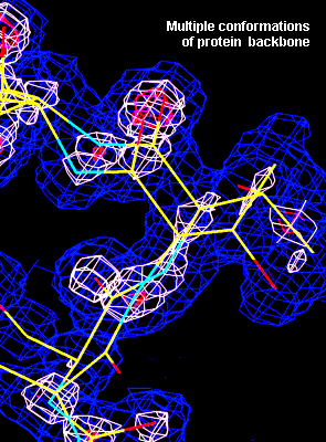
Figure 1. Left panel : At atomic resolution, multiple conformations of the protein side chains and of the protein backbone can be traced. At lower resolution, only an average structure can be fitted and the resulting model may not be a correct representation of the structure.

Figure 2 (above) shows an example for correlated multiple conformations in the molecule : In the centre of the panel is the PMS molecule which is bound to Ser216 (not shown). If PMS assumes the conformation with the Phenyl ring to the right, the distance to the His62 N(E2) is too close. In this case, the His62 ring will be in the lower of the two positions. At the same time, the position of the N of Asn153 will correlate with the sulfate oxygen on the PMS. Analysis of occupation numbers and the hydrogen bond geometry show that the upper conformation of Asn153 coexists with the PMS conformation showing the Phenyl ring on the right side.
Finally, the electron density of a Tryptophane 235, showing the holes in the rings. Such pretty pictures are often used to illustrate the quality of a high resolution refinement of a protein structure.
Figure 3 : Electron density detail of Trp235 demonstrating the high quality of the refined gg36 structure.
In order to obtain a high resolution refinement representing the features of the structure as accurately as possible, anisotropic temperature factors have to be used and Fiedel opposites in the data set must not be merged. Another very important feature is that the data were obtained from one flash-cooled single crystal to avoid merging of different data sets. The fact that we have data of extraordinary quality also allows us determine the position of hydrogen atoms.
Finding Hydrogen atoms in
atomic resolution protein refinements
The scattering of H atoms is very weak, and falls off rapidly
with increasing diffraction angle. As a result, it is difficult
to find H atoms in x-ray experiments. With data to atomic
resolution (better than about 1.2 A), however, it is possible to
locate H atoms in protein structures. Since the H scattering is
mostly confined to only moderate resolution, why is it that high
resolution is needed ? It appears that the calculation of
difference maps using a model refined with normal weights
introduces a lot of noise into the maps because the coordinates
of the protein 'heavy' atoms (C,N,O) are biased by contributions
from the valence electrons. This can be accounted for to some
extent by artificially up-weighting the higher angle reflections.
The least-squares fitted x,y,z then more closely approximate the
true nuclear positions, and difference maps are correspondingly
less biased, and H atoms are more visible. In the electron
density maps shown in figures 4 and 5, the protein model (without
H) was refined with enhanced weights for higher angle
reflections. The protein 'heavies' are contoured at 3 sigma
(2Fo-Fc) using all the data from 17-0.99 A. The H positions
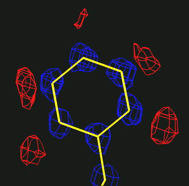
(density in red) are clear in an Fo-Fc map contoured at 2.5 sigma
using data from 17-1.25 A.

Fig.4 : Electron density
map showing the hydrogen atoms of a Phenylalanine residue.

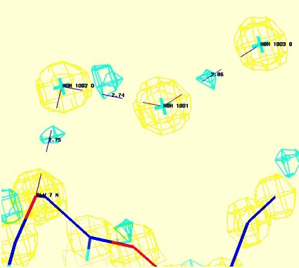
Figure 5 : Electron density map showing Hydrogen bond network. Cyan contours represent hydrogen atoms.
![]() Back to X-ray Facility Introduction
Back to X-ray Facility Introduction
LLNL Disclaimer
This World Wide Web site conceived and maintained by
Bernhard Rupp (br@llnl.gov)
Last revised April 06, 1999 11:03
UCRL-MI-125269